Ventral and dorsal exterior view of heart? Instant anatomy is a specialised web site for you to learn all about human anatomy of the body with diagrams, podcasts and revision questions ventral exterior view of the heart.
Ventral Exterior View Of The Heart, Point to any region of the large image, that region will then be highlighted in the smaller image to the left to help you locate it. Internal and external view of the developing head and neck at week 4. The ventral view of internal structure of heart shows two auricles, one ventricle, truncus arteriosus and the valves to keep the blood flowing in one direction.
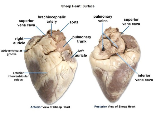 Ventral View Of Sheep Heart Labeled Pictures to Pin on From pinsdaddy.com
Finally learn the great vessels of the heart and their relationships to one another. Ventral refers to the front side of an organ, or that part of the organ that is facing front. I usually learn the vessels from the view of blood circulating through the heart, starting.
Lay the heart on the dissecting tray, anterior surface up.
It features a large pulmonary trunk that extends off the top and auricle flaps that cover the atria. For more anatomy content please follow us and visit our website: The arrowed bulge in panel c is fated to contribute to the atrioventricular canal. The right ventricle faces forward toward the sternum which lies exterior to the heart, and so constitutes the ventral surface of the heart. The ventral view of internal structure of heart shows two auricles, one ventricle, truncus arteriosus and the valves to keep the blood flowing in one direction. When you view the heart in a radiograph, these borders will be obvious.
Another Article :

Valves b) this side of the heart receives oxygenated blood. Notice the anterior interventricular sulcus posterior interventricular sulcus posterior 4. Instant anatomy is a specialised web site for you to learn all about human anatomy of the body with diagrams, podcasts and revision questions The heart has four chambers, and most diagrams will show the heart as it is viewed from the ventral side. Ventral refers to the front side of an organ, or that part of the organ that is facing front. Operation of the Heart Valves.
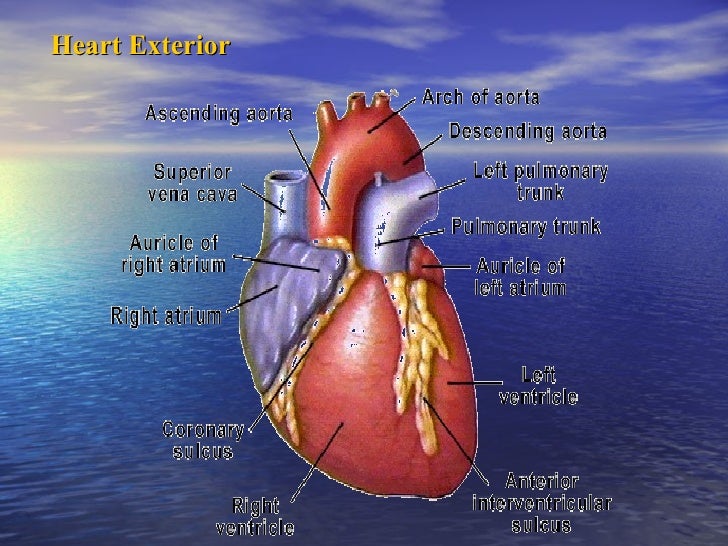
The ventral, or front, surface of the heart is distinguished by its curvature, whereas the backside is much flatter, notes shannan muskopf for the biology corner. Lay the heart on the dissecting tray, anterior surface up. Download scientific diagram | (top) dorsal and (bottom) ventral views of the heart of crocodylus porosus. The right ventricle faces forward toward the sternum which lies exterior to the heart, and so constitutes the ventral surface of the heart. Instant anatomy is a specialised web site for you to learn all about human anatomy of the body with diagrams, podcasts and revision questions The Heart And Great Vessels.

Ventral and dorsal exterior view of heart? Notice the anterior interventricular sulcus posterior interventricular sulcus posterior 4. The right ventricle faces forward toward the sternum which lies exterior to the heart, and so constitutes the ventral surface of the heart. A) the top two chambers of the heart are called atria. Part of the reason it is so difficult to learn is that the heart is not perfectly symmetrical, but it is so close that it becomes difficult to discern which side you are looking at (dorsel, ventral, left or right). Ansicht von ventral (vorne) des Herzens Stockfoto, Bild.

A) the top two chambers of the heart are called atria. The right ventricle faces forward toward the sternum which lies exterior to the heart, and so constitutes the ventral surface of the heart. The ventral, or front, surface of the heart is distinguished by its curvature, whereas the backside is much flatter, notes shannan muskopf for the biology corner. Internal and external view of the developing head and neck at week 4. Anatomynote.com found external heart anatomy anterior view from. front cardiovascular/blood anatomy Pinterest.

For more anatomy content please follow us and visit our website: The ventral, or front, surface of the heart is distinguished by its curvature, whereas the backside is much flatter, notes shannan muskopf for the biology corner. The right ventricle faces forward toward the sternum which lies exterior to the heart, and so constitutes the ventral surface of the heart. Ventral and dorsal exterior view of heart? Choose words from the list to complete the sentences below. gross anatomy of the heart anterior view Paramedic Study.
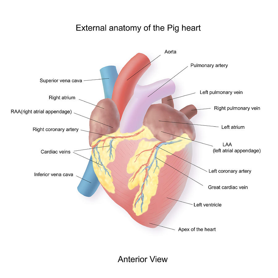
The arrowed bulge in panel c is fated to contribute to the atrioventricular canal. When you view the heart in a radiograph, these borders will be obvious. We are pleased to provide you with the picture named external heart anatomy anterior view.we hope this picture external heart anatomy anterior view can help you study and research. Answer all labels on the space provided. Finally learn the great vessels of the heart and their relationships to one another. Pig Heart Drawing at Explore.

B) these structures stop blood flowing backwards into the atria. Download scientific diagram | (top) dorsal and (bottom) ventral views of the heart of crocodylus porosus. Point to any region of the large image, that region will then be highlighted in the smaller image to the left to help you locate it. A) the top two chambers of the heart are called atria. When you view the heart in a radiograph, these borders will be obvious. Posterior heart surface Heart anatomy, Circulatory.
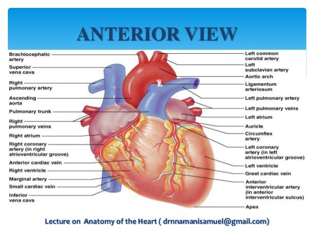
Ventral refers to the front side of an organ, or that part of the organ that is facing front. The front side of the heart is often identified by the coronary sinus that runs. The arrowed bulge in panel c is fated to contribute to the atrioventricular canal. The heart has four chambers, and most diagrams will show the heart as it is viewed from the ventral side. The heart dissection is probably one of the most difficult dissections you will do. Anatomy of the Heart.

Label the dorsal and ventral external features of the frog�s heart. I usually learn the vessels from the view of blood circulating through the heart, starting. Ventral refers to the front side of an organ, or that part of the organ that is facing front. The heart has four chambers, and most diagrams will show the heart as it is viewed from the ventral side. The ventral, or front, surface of the heart is distinguished by its curvature, whereas the backside is much flatter, notes shannan muskopf for the biology corner. Human Heart Anterior View Heart Anatomy Anatomy And.
Left hand side c) this is the largest artery in the body. The ventral view of internal structure of heart shows two auricles, one ventricle, truncus arteriosus and the valves to keep the blood flowing in one direction. Ventral refers to the front side of an organ, or that part of the organ that is facing front. (a) ventral view depicting the early heart, maxillary and mandibular processes of arch 1, arch 2, and the oropharyngeal. Point to any region of the large image, that region will then be highlighted in the smaller image to the left to help you locate it. What is the ventral surface of the heart? Quora.
![New Page 1 [classroom.sdmesa.edu] New Page 1 [classroom.sdmesa.edu]](http://classroom.sdmesa.edu/anatomy/images/Sheep_heart_labeled/Heart_arteries_labeled.jpg)
The ventral view of internal structure of heart shows two auricles, one ventricle, truncus arteriosus and the valves to keep the blood flowing in one direction. The heart has four chambers, and most diagrams will show the heart as it is viewed from the ventral side. Part of the reason it is so difficult to learn is that the heart is not perfectly symmetrical, but it is so close that it becomes difficult to discern which side you are looking at (dorsel, ventral, left or right). This means that as you look at the heart, the left side refers to the patient�s left side and not your left side. Notice the anterior interventricular sulcus posterior interventricular sulcus posterior 4. New Page 1 [classroom.sdmesa.edu].

Left hand side c) this is the largest artery in the body. Instant anatomy is a specialised web site for you to learn all about human anatomy of the body with diagrams, podcasts and revision questions The heart has four chambers, and most diagrams will show the heart as it is viewed from the ventral side. Left hand side c) this is the largest artery in the body. The right ventricle faces forward toward the sternum which lies exterior to the heart, and so constitutes the ventral surface of the heart. Heart Anatomy Tutorial YouTube.

The heart has four chambers, and most diagrams will show the heart as it is viewed from the ventral side. When you view the heart in a radiograph, these borders will be obvious. Notice the anterior interventricular sulcus posterior interventricular sulcus posterior 4. Distinguish the four chambers of the heart. We are pleased to provide you with the picture named external heart anatomy anterior view.we hope this picture external heart anatomy anterior view can help you study and research. Ventral View Heart.

Distinguish the four chambers of the heart. Ventral refers to the front side of an organ, or that part of the organ that is facing front. (a) ventral view depicting the early heart, maxillary and mandibular processes of arch 1, arch 2, and the oropharyngeal. The arrowed bulge in panel c is fated to contribute to the atrioventricular canal. For more anatomy content please follow us and visit our website: Anterior External View of Heart.

Left hand side c) this is the largest artery in the body. Valves b) this side of the heart receives oxygenated blood. Notice the posterior (dorsal) view: Choose words from the list to complete the sentences below. Distinguish the four chambers of the heart. Ventral View Of Sheep Heart Labeled Pictures to Pin on.


