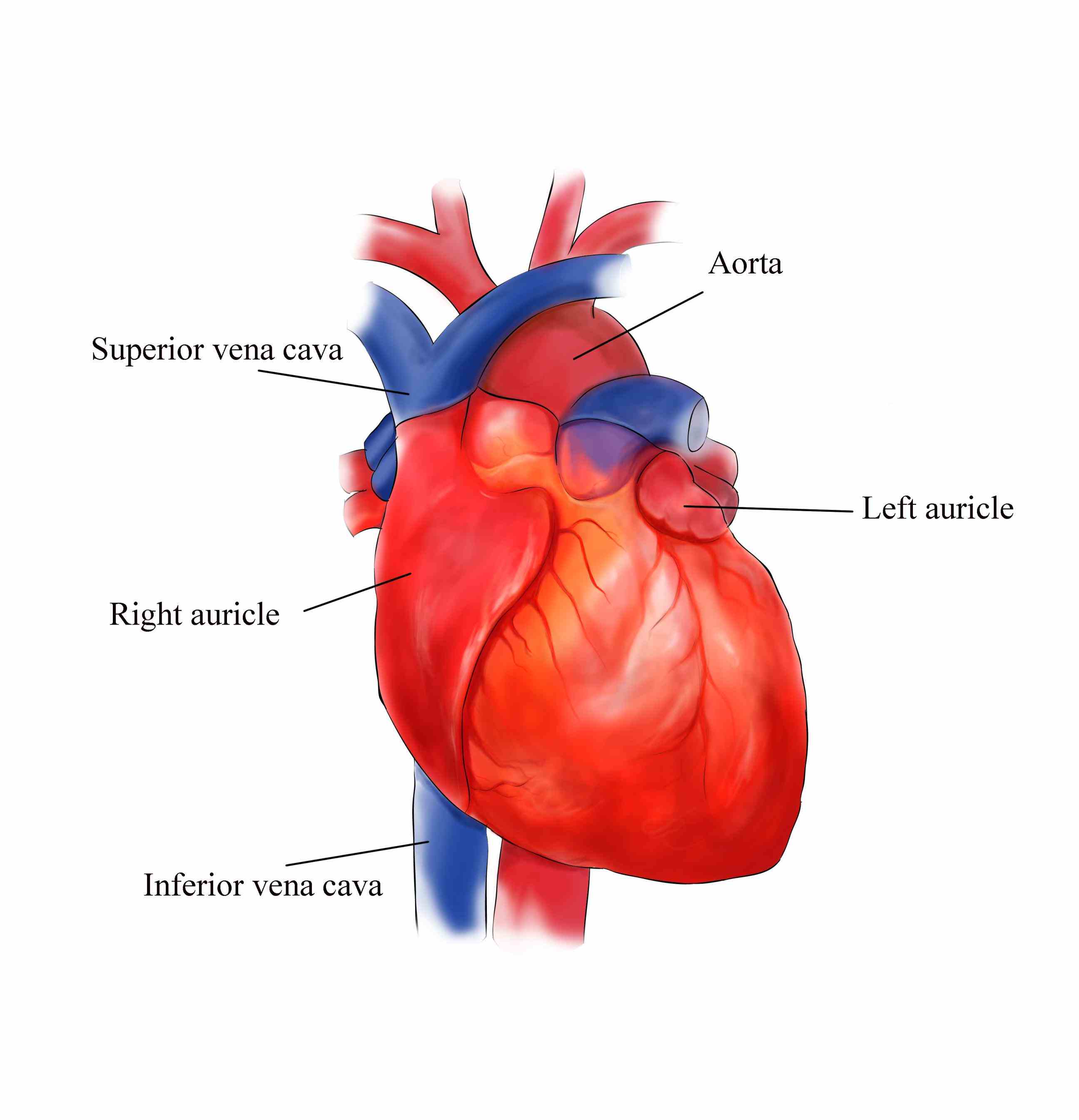
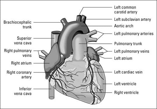
The coronary vessels are responsible for the delivery of blood to the heart muscle, or myocardium. Containing cholesterol, resulting in coronary heart disease. exterior view of the heart.
Exterior View Of The Heart, Layers of the pericardium 1. For more anatomy content please follow us and visit our website: This online quiz is called external anterior view of heart
 External Structure Of Heart Anatomy Diagram From medicinebtg.com
The vessels colored blue indicate the transport of blood with relatively low content of oxygen and high content of carbon dioxide. Myocardium, the thick middle layer of muscle that allows your heart chambers to contract and relax to pump blood to your body. 5th to 8th thoracic vertebrae.
Endocardium, the thin inner lining of the heart chambers that also forms the surface of the valves.
The light blue arrows show that blood enters the right atrium of your heart from the superior and inferior vena cavae. Also note the three borders of the heart: Left hand side c) this is the largest artery in the body. Right border (1) made up of the right atrium inferior border (2) made up of right atrium, right ventricle and left ventricle left border (3) made up of the left ventricle when you view the heart in a radiograph, these borders will be obvious. The heart appears trapezoid in the posterior and anterior views. Superior view of sheep heart:
Another Article :

Myocardium, the thick middle layer of muscle that allows your heart chambers to contract and relax to pump blood to your body. Health information and tools > patient care handouts > external view of the heart: We are pleased to provide you with the picture named external heart anatomy anterior view.we hope this picture external heart anatomy anterior view can help you study and research. License image this cross section of the heart shows the right ventricle, tricuspid valve, left ventricle, bicuspid (mitral) valve, left atrium, right atrium, superior vena cava, inferior vena cava, aorta, aortic valve, papillary muscle, chordae tendineae, and trabeculae carneae. The surface projections of the heart represent points on the thoracic wall that map out the outline and valves of the heart. Anatomy of the Heart Diagram View.

Right border (1) made up of the right atrium inferior border (2) made up of right atrium, right ventricle and left ventricle left border (3) made up of the left ventricle when you view the heart in a radiograph, these borders will be obvious. This is an online quiz called anatomy of the heart (external view) there is a printable worksheet available for download here so you can take the quiz with pen and paper. External features of the heart 1. A) the top two chambers of the heart are called atria. The heart ventricular walls consist of three layers: Operation of the Heart Valves.

The annotations reflect general features of the heart. Those coronary vessels which run along the surface of the heart are known as. We are pleased to provide you with the picture named external heart anatomy anterior view.we hope this picture external heart anatomy anterior view can help you study and research. Watch this video to learn about the ted (technology, entertainment, design) conference held in march 2011. Choose words from the list to complete the sentences below. Dr.Y.M.Kadiyani External Heart Anatomy.

The external surface of the heart is notable for 3 main sulci (grooves): Containing cholesterol, resulting in coronary heart disease. The surface projections of the heart represent points on the thoracic wall that map out the outline and valves of the heart. External view in question 1. If you click your left mouse button, the name of that structure will appear to identify it. לב האדם ויקיפדיה.

External view of the heart: The heart ventricular walls consist of three layers: The heart appears trapezoid in the posterior and anterior views. External view in question 1. The 1.epicardium, the 2.myocardium (cardiac. 1 Heart anatomy from the anterior view (left) and.

If you click your left mouse button, the name of that structure will appear to identify it. The 1.epicardium, the 2.myocardium (cardiac. The heart appears trapezoid in the posterior and anterior views. For more anatomy content please follow us and visit our website: For optimal viewing of this interactive, view at your screen’s default zoom setting (100%) and with your browser window view maximised. 2 the heart.

The light blue arrows show that blood enters the right atrium of your heart from the superior and inferior vena cavae. External features of the heart 1. Cardiac veins, on the other hand, return deoxygenated blood to the lungs. The model was created from scratch in zbrush. Health information and tools > patient care handouts > external view of the heart: Illustration Of Human Heart, Front View Digital Art by.

Pericardium pericardium is a fibroserous sac that encloses the heart and roots of the great vessels. Point to any region of the large image, that region will then be highlighted in the smaller image to the left to help you locate it. If you click your left mouse button, the name of that structure will appear to identify it. From the right ventricle, blood is pumped to your lungs through the pulmonary arteries. Arteries carry blood away from the heart while veins carry blood into the heart. External Structure Of Heart Anatomy Diagram.
Also note the three borders of the heart: Left hand side c) this is the largest artery in the body. External view in question 1. Body of sternum 2nd to 6th costal cartilages posterior: Pericardium pericardium is a fibroserous sac that encloses the heart and roots of the great vessels. Anatomy And Physiology Of Heart Quiz Anatomy Drawing Diagram.

This online quiz is called external anterior view of heart This online quiz is called external anterior view of heart B) these structures stop blood flowing backwards into the atria. Superior view of sheep heart: Watch this video to learn about the ted (technology, entertainment, design) conference held in march 2011. anterior heart Quiz.

If you click your left mouse button, the name of that region will appear to identify it. The coronary vessels are responsible for the delivery of blood to the heart muscle, or myocardium. Instant anatomy is a specialised web site for you to learn all about human anatomy of the body with diagrams, podcasts and revision questions Body of sternum 2nd to 6th costal cartilages posterior: Those coronary vessels which run along the surface of the heart are known as. Heart Anatomy Tutorial YouTube.

This online quiz is called external anterior view of heart Health information and tools > patient care handouts > external view of the heart: For optimal viewing of this interactive, view at your screen’s default zoom setting (100%) and with your browser window view maximised. Learn with this interactive gamee the components of the heart. Heart, external view (normal) personal ranking imformation statistics ranking descripción. External heart anatomy anterior view.

Also note the three borders of the heart: Photo about afternoon exterior view of the basilica of the sacred heart of paris, france. This model shows the external anatomy of the human heart. Anatomynote.com found external heart anatomy anterior view from. The 1.epicardium, the 2.myocardium (cardiac. CLASS BLOG BIO 202 Heart Anatomy.

Photo about afternoon exterior view of the basilica of the sacred heart of paris, france. Anatomy of the heart dr. Pericardium, the sac that surrounds your heart. License image this cross section of the heart shows the right ventricle, tricuspid valve, left ventricle, bicuspid (mitral) valve, left atrium, right atrium, superior vena cava, inferior vena cava, aorta, aortic valve, papillary muscle, chordae tendineae, and trabeculae carneae. The coronary vessels are responsible for the delivery of blood to the heart muscle, or myocardium. Science Source Pig Heart, Exterior Anatomy.

There have never been sufficient kidney donations to provide a kidney to each person needing one. External features of the heart 1. If you click your left mouse button, the name of that structure will appear to identify it. A) the top two chambers of the heart are called atria. The outer layer of the heart wall is the epicardium, the middle layer is the myocardium, and the inner layer is the endocardium. Figuring Out Cardiac Anatomy Your Heart dummies.


